
Introduction
Folate—vitamin B9—is not just a prenatal supplement. In its active form, 5-methyltetrahydrofolate (5-MTHF), it is an essential cofactor for nucleotide synthesis, methylation reactions, and neurotransmitter production. The brain is especially dependent on an uninterrupted supply of 5-MTHF. Yet, in a substantial subset of individuals with autism spectrum disorder (ASD), the brain is starved of folate even when blood folate levels are normal – a condition known as cerebral folate deficiency syndrome (CFDS).
A growing body of evidence points to a specific, self-reinforcing mechanism at the blood–brain barrier (BBB) that links mitochondrial energy failure to impaired folate transport, which in turn further damages mitochondria. This is the ATP–folate transport vicious cycle—a conceptual framework that explains how a transient or genetic vulnerability can spiral into a chronic neurodevelopmental and neuropsychiatric conditions.
The Players: ATP, Folate, and the Folate Receptor Alpha (FRα)
To understand the cycle, one must first appreciate the unique logistics of folate delivery to the central nervous system (CNS).
Folate cannot simply diffuse into the brain. It must be actively transported across the choroid plexus epithelium, the interface between blood and cerebrospinal fluid (CSF). The primary route is receptor-mediated endocytosis via the folate receptor alpha (FRα). FRα binds 5-MTHF with high affinity and internalizes it through a process that is strictly ATP-dependent. In other words, getting folate into the brain takes a certain amount of energy.
Alternately, a secondary route exists: the reduced folate carrier (RFC). This is important. However, RFC has a low affinity for folate at the BBB and is not normally the dominant transporter for CNS folate delivery. Its capacity is limited to compensate when FRα function is completely compromised.
ATP (adenosine triphosphate) is the universal energy currency of the cell, produced predominantly by mitochondria through oxidative phosphorylation. The brain is one of the most energy-demanding organs in the body. When mitochondrial function falters – as it does in a significant fraction of ASD cases – ATP production declines.
The Vicious Cycle:
The ATP–folate transport cycle can be broken down into a feed-forward loop with three interconnected nodes.
Node 1: Mitochondrial dysfunction → ATP depletion.
Mitochondrial dysfunction is a well-replicated biological abnormality in ASD. Studies suggest that between 30–50% of children with ASD possess biomarkers consistent with mitochondrial dysfunction, and many show reduced activity in electron transport chain complexes. When mitochondria fail, ATP availability at the choroid plexus falls. Mitochondrial function may be analyzed with MitoSwab™, a noninvasive cheek swab that can detail mitochondrial performance.
Node 2: ATP depletion → impaired FRα-mediated folate transport → cerebral folate deficiency.
Because FRα-mediated endocytosis is ATP-dependent, energy failure directly throttles folate entry into the CNS. This mechanism is not merely theoretical. In a landmark clinical observation, a child with ASD and mitochondrial disease was found to have CFD without FRα autoantibodies, leading the authors to conclude that lack of ATP availability contributing to mitochondrial dysfunction resulted in the impaired transportation of 5-MTHF into the CNS. The same logic has been extended to Kearns–Sayre syndrome, where failed ATP production explains low CSF 5-MTHF.
Node 3: Folate deficiency → further mitochondrial damage → deeper ATP depletion.
Folate is not merely a passenger in this system; it is essential for mitochondrial integrity. 5-MTHF is required for the synthesis of S-adenosylmethionine (SAM), the principal methyl donor for DNA, RNA, proteins, and lipids. SAM-dependent methylation regulates the expression of genes critical for mitochondrial biogenesis and function. Folate also supports the one-carbon metabolism (OCM) network, which supplies methyl groups for the synthesis of coenzyme Q10, creatine, and other mitochondrial cofactors. When folate is deficient, OCM is depleted, and the mitochondrial machinery that depends on methylation and one-carbon supply becomes destabilized.
Children with ASD and mitochondrial dysfunction may be especially vulnerable to folate insufficiency: impaired mitochondria may produce insufficient ATP to support energy-dependent, FRα-mediated folate transport, thereby depleting one-carbon metabolism (OCM), which is needed to sustain mitochondrial function. Indeed, a vicious cycle.
A Parallel Aggravator: Folate Receptor Alpha Autoantibodies (FRAAs)
In a separate but convergent pathway, autoantibodies against FRα can directly block folate binding and impair FRα function. FRAAs are found in a strikingly high proportion of children with ASD – estimates range from 50% to 75% in some clinical cohorts, with systematic reviews attributing 83% of CFD cases in ASD to FRAAs. These antibodies can be blocking (preventing 5-MTHF from binding) or binding (interfering with receptor function through other mechanisms).
FRAAs and mitochondrial dysfunction are not mutually exclusive; they can coexist and synergize. A child with both FRAA-mediated blockade and mitochondrial ATP depletion faces a double hit to the same transport step. Furthermore, the immune activation that produces FRAAs may itself be linked to mitochondrial dysfunction and oxidative stress, creating another layer of reinforcement.
The ATP–folate cycle therefore operates both with and without FRAAs. In FRAA-negative children with ASD and mitochondrial dysfunction, the cycle is driven purely by energy failure. In FRAA-positive children, the cycle may be initiated or accelerated by antibody-mediated blockade, with mitochondrial compromise as a secondary amplifier.
Why This Matters for the Autistic Brain
The ATP–folate vicious cycle has profound consequences for neurodevelopment and function.
- Impaired methylation and gene regulation. Depleted SAM pools reduce methylation of DNA and histones, altering the expression of genes involved in synaptic plasticity, neuroinflammation, and mitochondrial function.
- Neurotransmitter imbalance. Folate is required for the synthesis of serotonin, dopamine, and norepinephrine. Cerebral folate deficiency sndrome can therefore contribute to the monoamine abnormalities frequently observed in ASD.
- Oxidative stress amplification. Folate deficiency impairs the regeneration of tetrahydrobiopterin (BH4) and the production of glutathione, the brain’s master antioxidant. Mitochondria, already compromised, become more vulnerable to oxidative damage, further suppressing ATP production.
- Clinical phenotype. CFD in ASD has been associated with more severe neurological deficits, including low-functioning autism with neurological abnormalities, motor dysfunction, and communication impairments. The cycle may also help explain the transgenerational patterns observed with FRAAs and the variable severity of the autism phenotype.
Breaking the Cycle: Therapeutic Implications
The ATP–folate vicious cycle is not a closed loop without exit points. Understanding its nodes reveals therapeutic strategies.
- Leucovorin (folinic acid): This reduced form of folate can bypass the blocked FRα by using the reduced folate carrier and other transport mechanisms, delivering folate to the CNS even when FRα function is impaired. Clinical trials have shown that leucovorin improves symptoms in children with ASD and CFD, particularly in those who are FRAA-positive. Notably, leucovorin has also been associated with improved mitochondrial function – higher complex I activity and increased maximal respiratory capacity—suggesting that restoring folate can help break the cycle at Node 3.
- Targeting mitochondria: Interventions that support mitochondrial function—such as coenzyme Q10, carnitine, B-vitamins, and antioxidants—may help maintain ATP production at the choroid plexus, preserving FRα-mediated folate transport even in the absence of folate supplementation. A small study conducted by Dr. Richard Frye showed promise in regard to this. However, these approaches require rigorous clinical validation in ASD. A small study
- Screening and stratification: The cycle framework argues for biomarker-guided stratification. Children with ASD and evidence of mitochondrial dysfunction (e.g., elevated lactate, reduced respiratory chain activity) or FRAA positivity may be the most likely to benefit from early folate-targeted interventions. Cerebral folate deficiency can be identified by measuring 5-MTHF in CSF via lumbar puncture, though a less invasive biomarker (FRAT® test that identifies and measures FRAA titers) is being explored.
- Early intervention: The cycle is likely most damaging during critical developmental windows. The observation that younger children respond more robustly to leucovorin than older children underscores the importance of early identification and intervention. With this is mind it is recommended that physicians become familiar with the FRAT® test and MitoSwab™.
A Unifying Lens on a Heterogeneous Condition
Autism is notoriously heterogeneous, and no single mechanism explains all cases. The ATP–folate transport vicious cycle does not claim to be a universal explanation. But it offers a mechanistically plausible, testable, and clinically actionable framework for a substantial subgroup of individuals—those with mitochondrial dysfunction, FRAAs, or both.
The cycle’s elegance lies in its feed-forward logic: a modest initial hit to energy production or folate transport can, over time, become a self-sustaining pathology. It bridges two previously separate literatures—mitochondrial dysfunction in ASD and cerebral folate deficiency—into a single dynamic model. And it makes a clear prediction: interventions that restore either ATP or folate availability may interrupt the cycle and yield clinical benefit.
For families and clinicians, the practical takeaway is that folate status in the brain is not the same as folate status in the blood. A child with normal serum folate can still have a brain starving for methylfolate if the energy-dependent transport machinery is failing. Recognizing the ATP–folate cycle as a distinct pathophysiological pathway is a step toward more precise, mechanism-based care in autism.
Did You Know? Folate Receptor Autoantibodies (FRAAs) may impede proper folate transport.
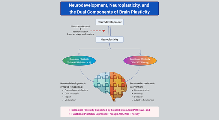
Folate (vitamin B9) is very important for your child’s brain development!
During pregnancy, it helps prevent neural tube defects and plays a big role in forming a normal and healthy baby’s brain and spinal cord. Folate also helps cells divide and assists in both DNA and RNA synthesis.
Emerging research suggests that the presence of FRAAs negatively impacts folate transport into the brain.
- Recent studies reveal that a large subgroup of children with autism spectrum disorder (ASD) have FRAAs.
- This suggests that a possible disruption in folate transport across the blood-cerebrospinal fluid (CSF) barrier may potentially influence ASD-linked brain development.
- Screening for the FRAAs in your child should be part of your early intervention strategies.
Is there a test for identifying Folate Receptor Autoantibodies (FRAAs)?
Yes, there is a test – The Folate Receptor Antibody Test (FRAT®) has emerged as a diagnostic tool for detecting the presence of FRAAs.
It is important to screen at an early age or as soon as possible as there may be corrective measures available. Please consult your physician for further information.
To request a test kit, click on the button below.

For information on autism monitoring, screening and testing please read our blog.
References
- https://www.mdpi.com/1422-0067/27/20/9101/pdf?version=1791968089#4#6#4
- https://pmc.ncbi.nlm.nih.gov/articles/PMC13304352/#1
- https://www.sciencedirect.com/science/article/pii/S1071909120300401
- https://www.frontiersin.org/journals/physiology/articles/10.3389/fphys.2014.00150/full
- https://www.mdpi.com/1422-0067/26/6/2479
- https://www.fratnow.com/pdf/Cerebral-Folate-Deficiency-Syndrome-early-diagnosis-intervention-and-treatment-strategies.pdf
- https://www.fratnow.com/pdf/Folinic-Acid-Improves-Verbal-Communication-in-Children-with-Autism-R.Frye-clinical-trial.pdf
- https://www.fratnow.com/pdf/Folate-Receptor-Alpha-Autoantibodies-in-ASD-diagnosis-treatment-prevention.pdf