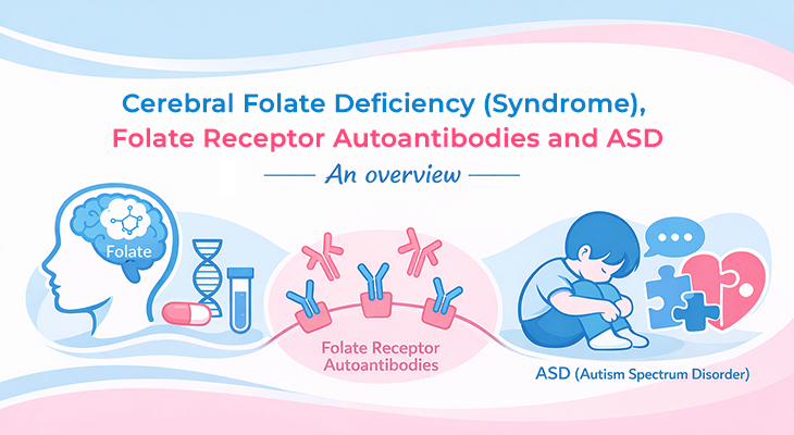

Folate (vitamin B9) is essential for brain development, neurotransmitter function, and neurological health. Yet many people with adequate folate in their blood still suffer from functional folate deficiency in the brain — because autoantibodies are blocking the transport system that delivers folate across the blood-brain barrier. These autoantibodies come in two distinct types: blocking and binding. Understanding the difference is critical, because failing to test for both can mean missing a diagnosis that has a straightforward treatment.
What Are Folate Receptor Autoantibodies?
Folate receptor alpha (FRα) is the primary transporter that carries 5-methyltetrahydrofolate (5-MTHF), the active form of folate, across the blood-brain barrier and into the cerebrospinal fluid (CSF). Folate receptor autoantibodies (FRAAs) are antibodies produced by the immune system that mistakenly target and bind to FRα, disrupting this transport process.
This disruption creates a condition called cerebral folate deficiency syndrome (CFDS) – low folate in the brain despite normal or even elevated serum folate levels. The consequences can be severe: neurodevelopmental delays, autism spectrum disorder, schizophrenia, depression, and neural tube defects during pregnancy.
There are two distinct types of FRAAs, each with a different mechanism of harm—and each requires a separate assay to detect.
Blocking Autoantibodies: The Direct Inhibitors
Blocking autoantibodies work through direct, competitive inhibition. They attach to the folate receptor at or near the folate-binding site, physically preventing folate from docking with the receptor and being transported into the cell.
This mechanism was elegantly demonstrated in a landmark study published in the New England Journal of Medicine, which found that serum from women who had pregnancies complicated by neural tube defects contained autoantibodies that blocked the binding of radiolabeled folic acid to folate receptors on placental membranes and inhibited folate uptake by cells. The autoantibodies displayed high binding affinity, explaining their potent blocking effect.
The clinical significance of blocking antibodies is substantial. Research has shown that subfertility risk was 12 times higher in women with blocking FR autoantibodies compared to those without them. In children with autism, blocking FRAAs have been associated with relatively better redox metabolism and inflammation markers, and better communication scores on adaptive behavior scales—suggesting that blocking and binding antibodies may define distinct clinical subgroups.
Binding Autoantibodies: The Indirect Disruptors
Binding autoantibodies operate through a different mechanism. Rather than directly occupying the folate-binding site, they attach to other parts of the receptor, altering its conformational structure and general position in the cell membrane. This change in receptor configuration impairs its ability to transport folate effectively.
But that is not the only consequence. Binding autoantibodies are also thought to trigger an antigen-antibody-mediated inflammatory response that further disrupts receptor function and contributes to the broader pathophysiology of folate transport disorders.
In a study of 94 children with autism spectrum disorder, children positive for binding FRAAs had significantly higher serum B12 levels compared to those negative for binding FRAAs. This finding suggests that binding antibodies may be associated with a distinct physiological profile that could serve as a biomarker for treatment response.
Why Testing for Both Types Is Essential
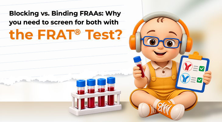
Here is the critical clinical point: blocking and binding autoantibodies can occur independently or together. Some patients have only blocking antibodies, some have only binding antibodies, and some have both. If a test only screens for one type, it will miss patients who are positive for the other.
The FRAT® (Folate Receptor Autoantibody Test), developed in the laboratory of Dr. Edward Quadros at SUNY Downstate, is the only assay available that screens for both blocking and binding folate receptor autoantibodies. It consists of two parts—a blocking assay and a binding assay—and reports separate titer levels for each type found.
The clinical rationale for testing both is straightforward: the downstream consequence of either antibody type is the same—impaired folate transport to the brain. But the physiological signatures differ. Research has shown that children positive for binding FRAAs have higher B12 levels, while children positive for blocking FRAAs have better redox and inflammation markers and better communication scores. These distinctions may have implications for treatment selection and monitoring.
Furthermore, autoantibody titers can fluctuate over time. FRAAs may be influenced by diet (particularly dairy exposure, given the homology between milk folate-binding proteins and human folate receptors) or by immune status at the time of testing. The NEJM study on cerebral folate deficiency syndrome speculated that autoantibody production could be induced by soluble folate-binding proteins in human or bovine milk, which share 91% amino acid sequence homology with membrane-bound folate receptors expressed on human choroid plexus epithelium.
Clinical Conditions Associated with FRAAs
Folate receptor autoantibodies have been documented in a range of conditions:
- Cerebral folate deficiency syndrome: Characterized by low CSF folate despite normal serum folate, with neurodevelopmental regression, seizures, and movement disorders.
- Autism spectrum disorder: FRAAs are prevalent and define a subgroup of children who may respond to folinic acid treatment.
- Neural tube defects: Blocking autoantibodies have been identified in 75% of women with affected pregnancies, a rate similar to the 70% reduction in NTDs achieved with periconceptional folic acid supplementation.
- Subfertility and miscarriage: Blocking autoantibodies are associated with a 12-fold increased risk of subfertility.
- Neuropsychiatric disorders: Depression, and schizophrenia have been linked to folate transport impairment.
The Treatment Implication
The good news is that identifying FRAAs—whether blocking, binding, or both—opens the door to targeted treatment. Folinic acid (leucovorin) and/or 5-MTHF can bypass autoantibody-blocked folate receptors and enter the cerebrospinal fluid via the reduced folate carrier, restoring folate levels within the central nervous system. This route of administration can ameliorate neuropsychiatric symptoms and support neurodevelopmental improvement.
Clinical trials have demonstrated that folinic acid improves verbal communication in children with autism and language impairment, and that binding FRAA may serve as a biomarker for leucovorin treatment response.
Conclusion
The distinction between blocking and binding folate receptor autoantibodies is not academic – it is clinically actionable. Blocking antibodies directly prevent folate from binding to its receptor, while binding antibodies alter receptor conformation and trigger inflammation. Both impair folate transport to the brain, but their physiological signatures differ, and they can occur independently or together.
The FRAT® test is the only available assay that detects both types, providing a complete picture of a patient’s folate transport status. For clinicians evaluating patients with unexplained neurodevelopmental, psychiatric, or reproductive concerns—particularly when serum folate appears normal—screening for both blocking and binding FRAAs is essential. Missing one type means missing the diagnosis. And missing the diagnosis means missing the opportunity to intervene with a treatment that can be genuinely transformative.
References:
- https://www.nejm.org/doi/10.1056/NEJMoa031145?url_ver=Z39.88-2003&rfr_id=ori:rid:crossref.org&rfr_dat=cr_pub%3dwww.ncbi.nlm.nih.gov#2
- https://pubmed.ncbi.nlm.nih.gov/27013943/#1
- https://www.nejm.org/doi/10.1056/NEJMoa043160?url_ver=Z39.88-2003&rfr_id=ori:rid:crossref.org&rfr_dat=cr_pub%20%200www.ncbi.nlm.nih.gov#2